

The Science
Helping the brain rewire itself, one movement at a time
A direct line to the brain
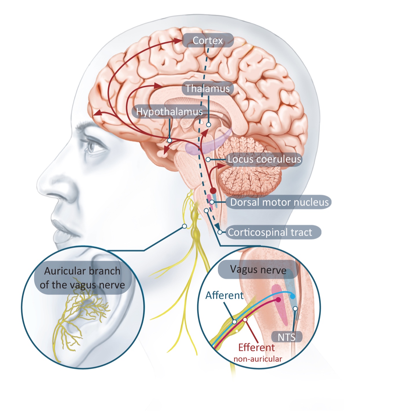
The vagus nerve is the longest nerve of its kind in the body, linking the brain to the heart, lungs and gut. What is less well known is that around 80% of its fibres carry signals up to the brain, making it one of the most powerful natural inputs we have for influencing how the brain works.
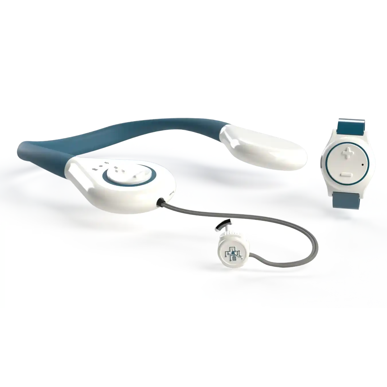
One small branch of the vagus nerve surfaces just under the skin of the outer ear. SmartVNS uses a comfortable earpiece to gently stimulate this branch. From there, signals travel up to the brainstem and on to the regions that release the brain's key learning chemicals.
Sources: Berthoud & Neuhuber, 2000 · Peuker & Filler, 2002
A "save" signal for the recovering brain
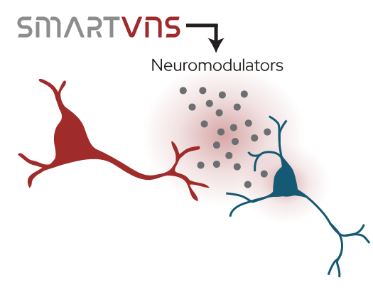
Every time we practise a movement, our neurons fire together and form short-lived connections. For those connections to stick, the brain needs a chemical signal that says "this moment mattered, keep it." Without that signal, most of what we practise fades away.
A brief pulse of vagus nerve stimulation releases exactly those chemicals (noradrenaline and acetylcholine) across the cortex. They don't carry the movement itself; they tell the rest of the brain that what just happened is worth remembering. In effect, SmartVNS hands the brain a save button at the very moment learning is most possible.
Sources: Hulsey et al., 2017 · Hays, 2016
What we can measure when stimulation is on
In healthy volunteers, we can directly observe the three changes that taVNS produces in the nervous system. Each one is a step in the chain from the ear to the muscle, and each one is amplified when stimulation is paired with movement.
Neuromodulators released
A short pulse of taVNS causes the pupils to dilate, a well-established sign that the brainstem has just released noradrenaline, the brain's "pay attention" chemical. The effect is consistent and stronger than a sham stimulation.
Cortex tunes in
EEG recordings over the motor cortex show a shift toward a more arousal- and learning-ready state during taVNS. The shift is largest exactly when the patient is moving, the moment that matters for recovery.
Brain-to-muscle pathway strengthens
Using Transcranial Magnetic Stimulation (TMS) to probe the corticospinal tract, we see that the pathway from motor cortex to muscle becomes significantly more excitable while stimulation is on, meaning the brain's signals reach the body more effectively during therapy.
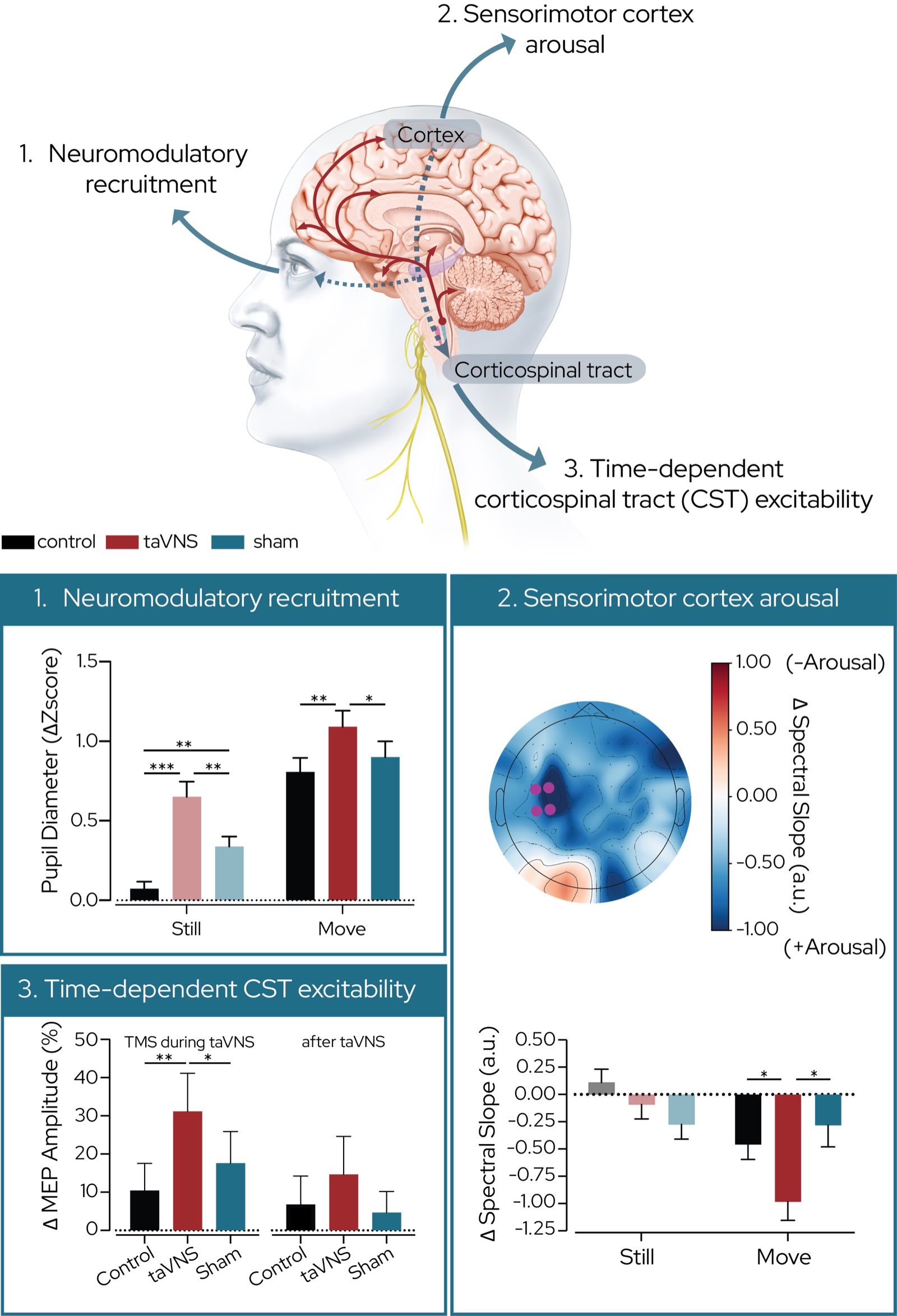
Source: Perrin et al., 2026
Paired to the movement that matters
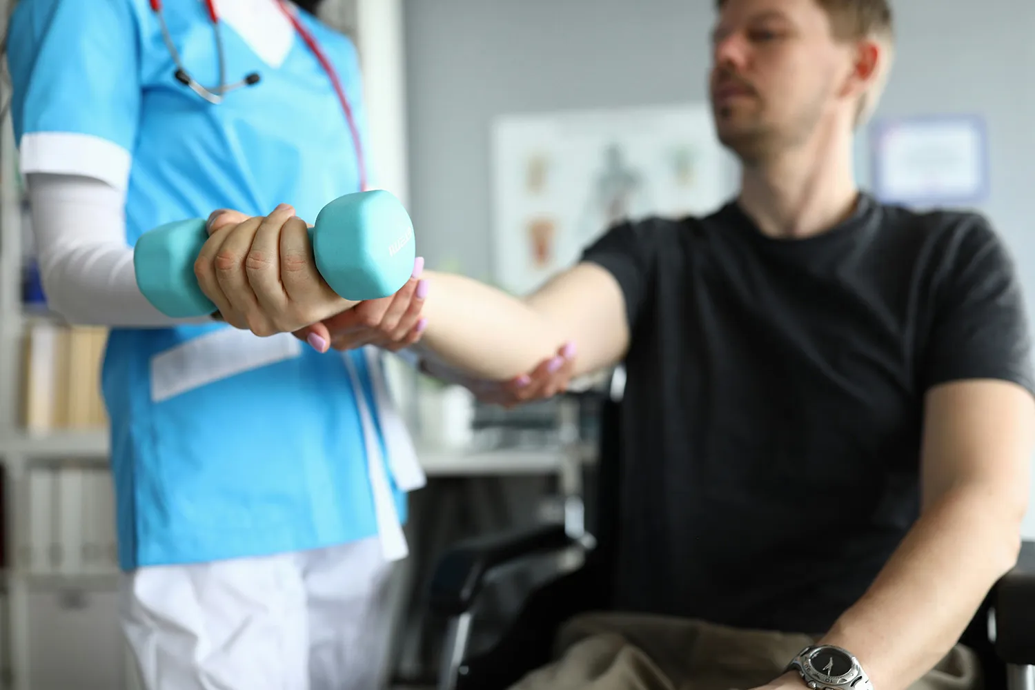
Stimulation alone is not enough. Research shows that pairing the stimulation with active, goal-directed movement is what drives lasting change in the brain's motor circuits.
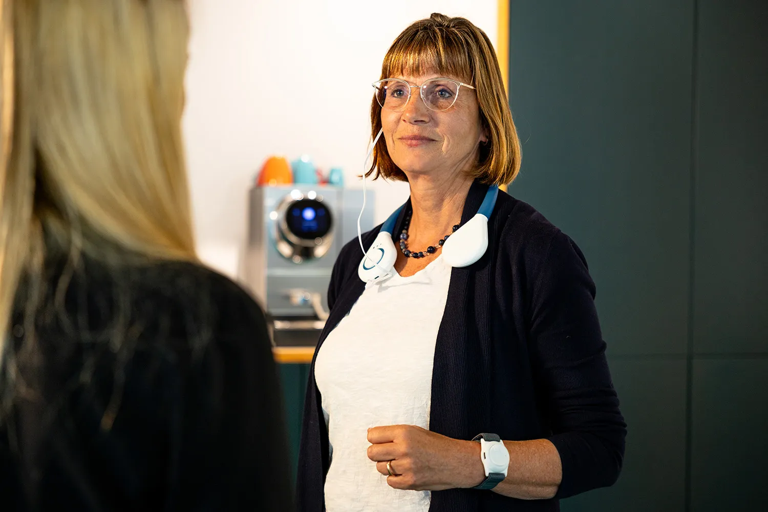
A wrist-worn motion sensor watches for the moments that matter (the patient reaching, lifting or grasping with their weaker arm) and triggers stimulation within a fraction of a second. This means each session reinforces recovery-relevant patterns, not the compensations that often emerge after a stroke.
Sources: Engineer et al., 2011 · Schambra & Hays, 2025

From principle to practice
The principle of pairing vagus nerve stimulation with rehabilitation is already established: in 2021 the US FDA approved an implanted version of this approach for chronic stroke recovery. SmartVNS is being developed to deliver the same biological mechanism non-invasively, through a comfortable earpiece, in a form that patients can use anywhere.
In our first clinical feasibility study, nine participants with stroke or spinal cord injury used SmartVNS across 20 therapy sessions over four weeks. The system delivered consistent, movement-paired stimulation across very different ability levels, was rated highly usable by both patients and therapists, and could be set up independently in under two minutes, even by patients with significant arm weakness. These early results lay the groundwork for the larger controlled trials now underway.
Sources: Dawson et al., 2021 · Lhoste et al., 2026
Our publications
Closed-Loop Movement-Paired Transcutaneous Auricular Vagus Nerve Stimulation for Upper-Limb Rehabilitation: a Feasibility Study
Read the open-access paper →
Published · 2026Transcutaneous auricular vagus nerve stimulation during movement selectively activates motor circuitry without additional cortical or autonomic effects
Read the paper →
Preprint · 2026Machine Learning-Based Real-Time Detection of Compensatory Trunk Movements Using Trunk-Wrist Inertial Measurement Units
Read the preprint →
Join Us
Join Skaaltec and help us transform rehabilitation by turning everyday activities into accessible therapy. Together, we can make recovery more effective and available to all.
Open Projects